
XRay Open Air MRI of CenLa
A hand X-ray is a black and white image that shows the inner structures of your hand, such as your bones and soft tissues. This diagnostic tool can help your doctor locate and understand.
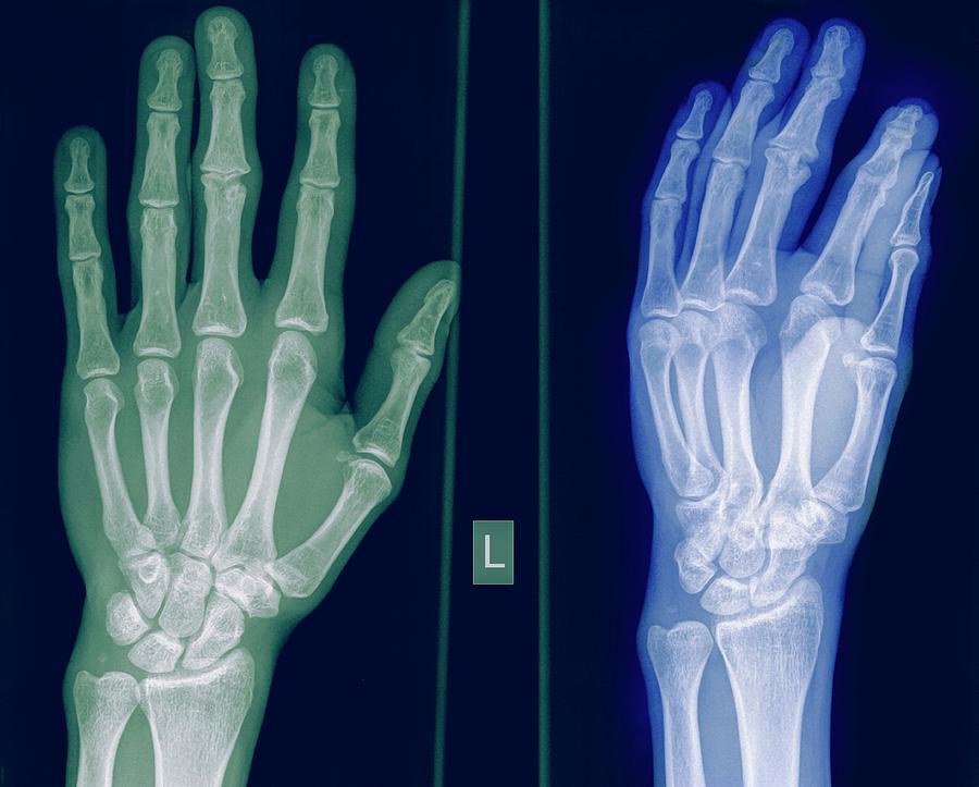
Hand XRay
1. Introduction Hand radiographs are frequently ordered as the first imaging modality in the assessment of patients presenting with peripheral arthritis. They can provide invaluable information about the bones, joints, mineralization, soft tissues and the distribution of abnormalities.

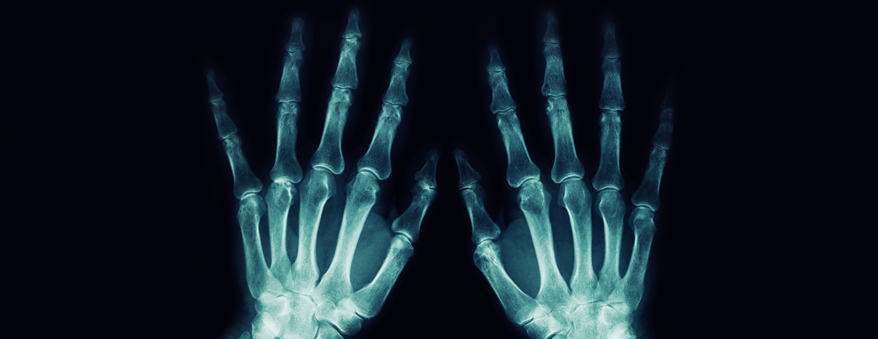
Xray of Both Human Hands.Normal Human Hands. Stock Image Image of inflammation, arthritis
Presentation Bilateral hand pain. Patient Data Age: 45 years Gender: Male x-ray bilateral and symmetrical involvement proximal interphalangeal joint space narrowing metacarpal heads erosions metacarpophalangeal joint space narrowing metacarpophalangeal joint osteopenia pancarpal and radiocarpal involvement with erosions carpometacarpal erosion
Xray Of A Healthy Hand Photograph by Photostockisrael Fine Art America
A hand x-ray is an imaging technique used to take pictures of hand bones using radiological waves known as "x" rays for medical purposes. Who do I need to see to get an x ray? If you have experienced an acute trauma to your hand, finger or wrist and think you may have an injury you should see your doctor.
Hand xray
Hand radiograph (an approach) Last revised by Mostafa El-Feky on 1 Oct 2020 Edit article Citation, DOI, disclosures and article data Hand radiographs are commonplace in the Emergency Department or the trauma reporting list. Systematic review

Hand xray. Causes, symptoms, treatment Hand xray
Key points. Finger injuries visible on X-ray include bone fractures, dislocations and avulsions. The hand comprises the metacarpal and phalangeal bones. Fractures and dislocations are usually straightforward to identify, so long as the potentially injured bone is fully visible in 2 planes. Finger joints commonly dislocate and are susceptible to.
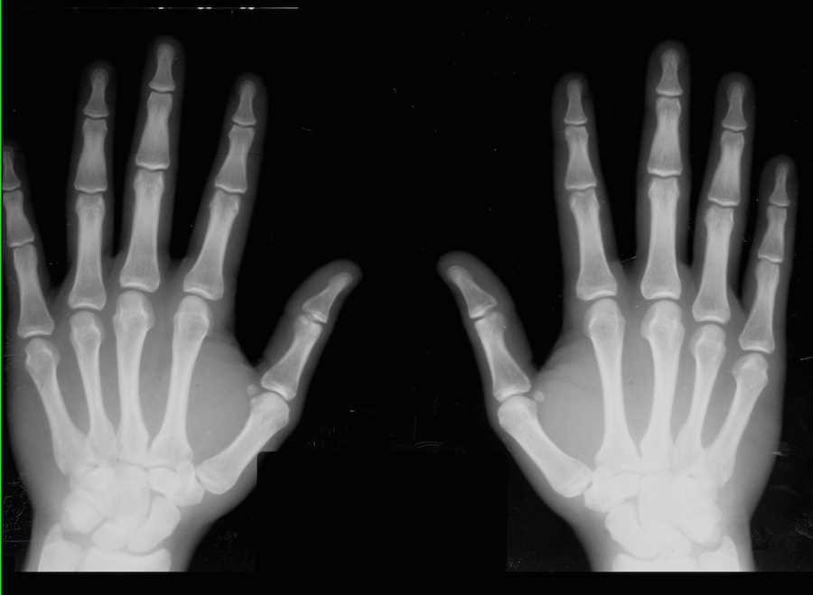
Normal Hands on Xray X Rays Case Studies CTisus CT Scanning
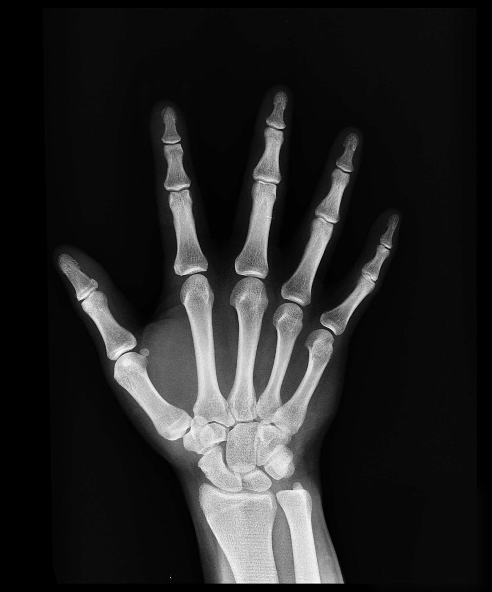
Shaft of third metacarpal. Neck of fifth metacarpal. Head of forth metacarpal. Metacarpophalangeal joint. Proximal phalanx. Middle phalanx. Distal phalanx. Sesamoid bones (flexor pollicis brevis, adductor pollicis). Terminal tuft.

Hand xray. Causes, symptoms, treatment Hand xray
X-ray - hand. How the Test is Performed. A hand x-ray is taken in a hospital radiology department or your health care provider's office by an x-ray technician. You will be asked to place your hand on the x-ray table, and keep it very still as the picture is being taken. You may need to change the position of your hand, so more images can be taken.
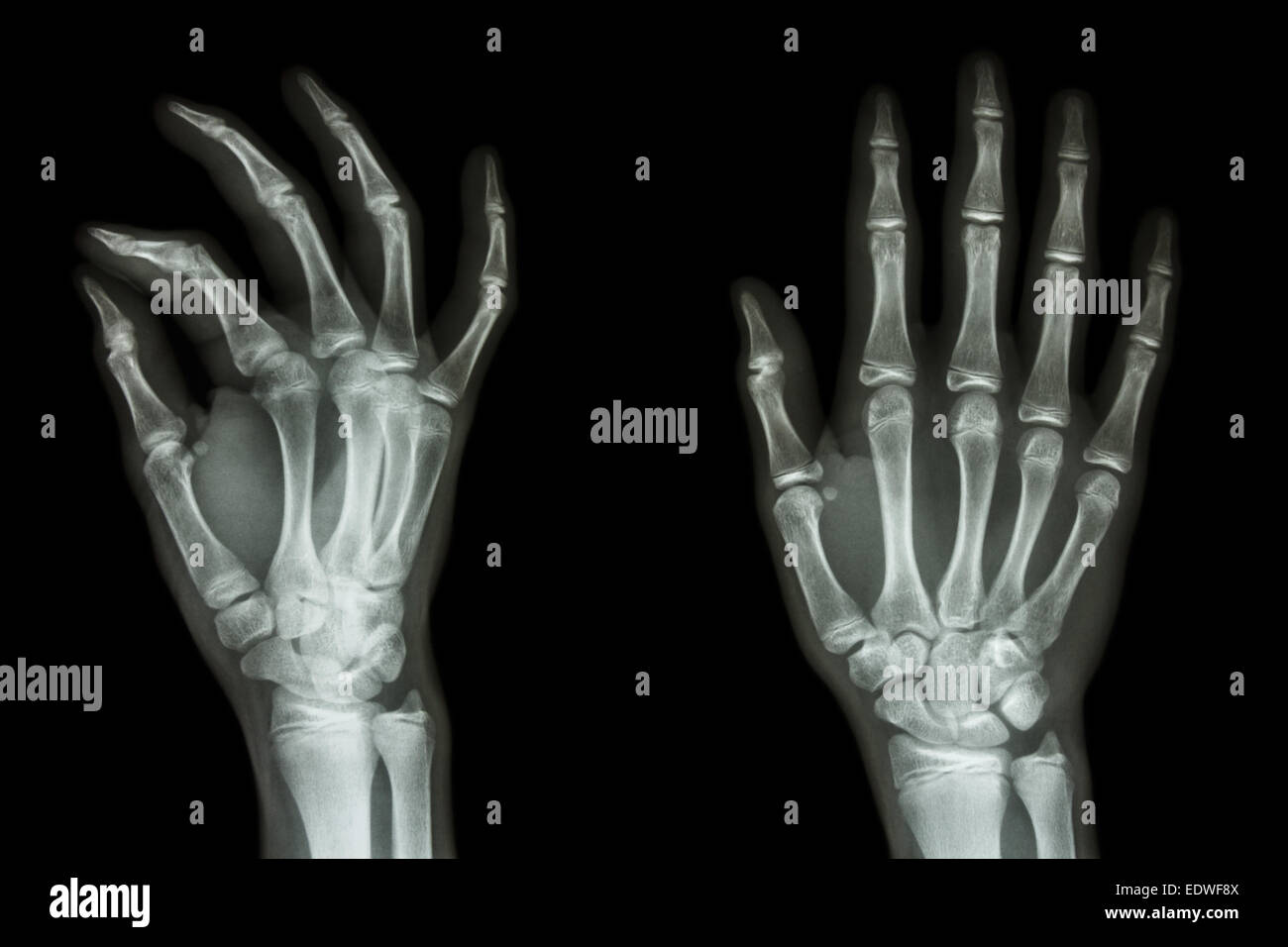
Xray Hand Normal High Resolution Stock Photography and Images Alamy
A hand X-ray (radiograph) is a test that creates a picture of the inside of your hand. The picture shows the inner structure ( anatomy) of your hand in black and white. Calcium in your bones absorbs more radiation, so your bones appear white on the X-ray.
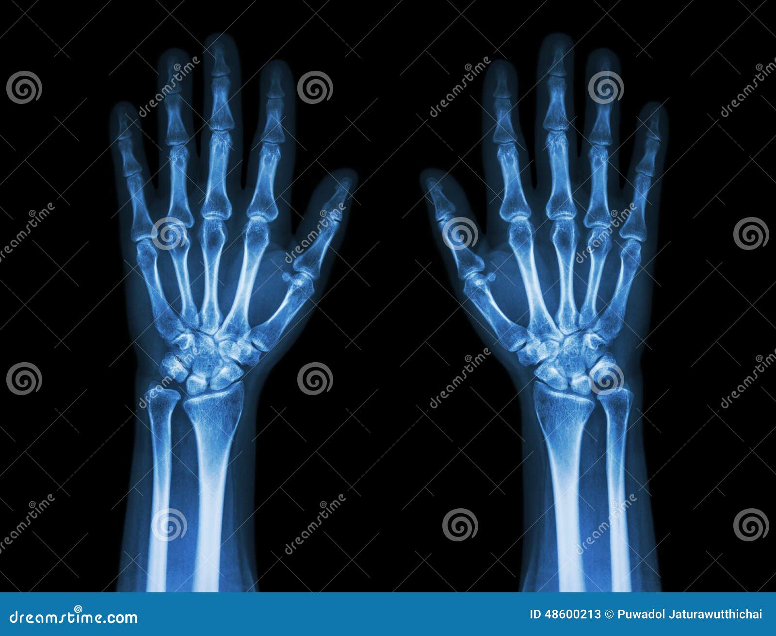
X Ray Hands Front View Normal Human Hands Stock Photos Free & RoyaltyFree Stock Photos from
Medical Encyclopedia → Hand x-ray Hand x-ray This test is an x-ray of one or both hands. How the Test is Performed A hand x-ray is taken in a hospital radiology department or your health care provider's office by an x-ray technician. You will be asked to place your hand on the x-ray table, and keep it very still as the picture is being taken.
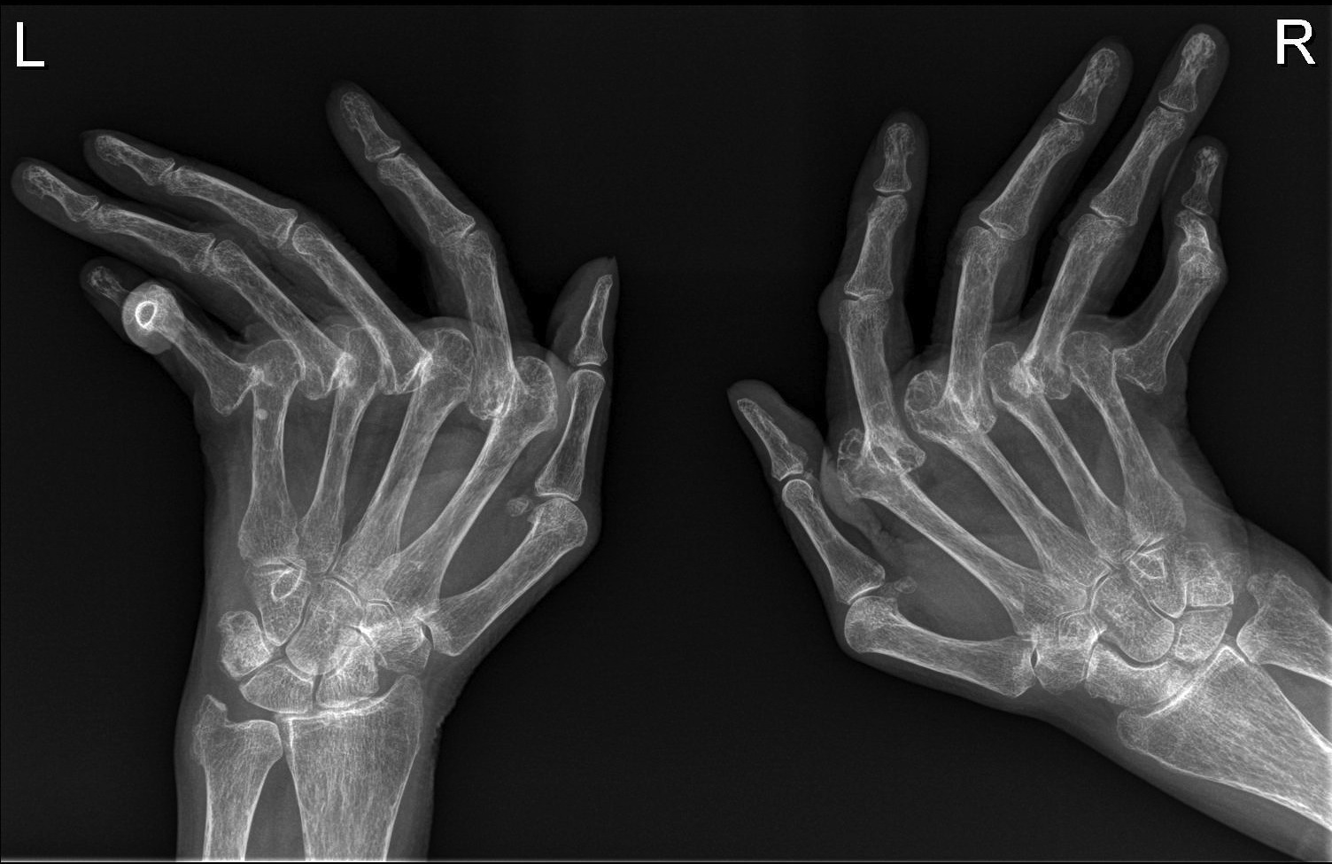
Rheumatoid arthritis hands Radiology at St. Vincent's University Hospital
POSTERIOR-ANTERIOR VIEW. This is the most commonly used view for interpretation. Finger deformities may not be noticed as patients are required to press their hands down firmly against the plate, while the X-Rays are shot from above.

Xray of an iodine dipped hand. Anatomy for artists, X ray, Hand anatomy
15) Your palm bones are hard to feel in your hand. Try looking at a real-life X-ray of the hand online. Your palm bones go from each finger to the wrist. Draw your 5 palm bones in ovals. -ray of a hand again to see the bones in your wrist. There are 8 small bones in your wrist. 17) Look at the X-ray to see the bones of your arm. Draw those 2 bones.

Xray of Hands Free Photo Download FreeImages
Hand x-ray is used to detect fractures, tumors, foreign objects, or degenerative conditions of the hand. Hand x-rays may also be done to find out a child's "bone age." This can help determine if a health problem is preventing the child from growing properly or how much growth is left. What Abnormal Results Mean Abnormal results may include:
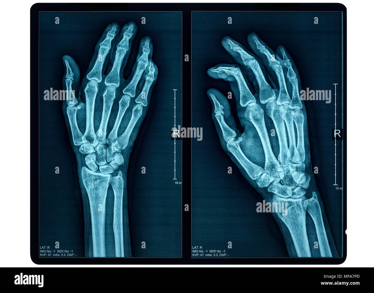
Hand Xray of an Adult Female Stock Photo Alamy
Hand x-rays are indicated for a variety of settings, including: trauma with suspected fracture suspected metacarpal dislocation foreign body detection and localization investigation of joint pain and/or deformity rheumatoid arthritis osteoarthrosis Projections Standard projections PA view
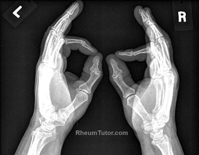
Approach to Hand XRays · RheumTutor
Access my FREE Online Membership today → https://www.thenotedanatomist.com___Unlock my Premium Tutoring Memberships → https://www.thenotedanatomist.com/premi.

Sports medicine stats Metacarpal fractures and other fractures of the hand Dr. David Geier
Hand series (summary) Last revised by Andrew Murphy on 23 Aug 2019 Edit article Citation, DOI, disclosures and article data This is a basic article for medical students and other non-radiologists A hand series (or hand x-ray) may be performed for a multitude of reasons.